- Main Page
- A1C Test
- Advance Directives
- Anxiety
- Aortic Aneurysm
- Aphrodisiacs
- Apple Cider Vinegar
- Arrhythmia
- Atrial Fibrillation - AFib
- Back Pain
- Blood Tests
- Blood Test Tubes
- Blood Types
- BMI Calculator
- Body Mass Index - BMI
- Bone Density Scan
- Bone Scan
- BPPV
- Bronchitis
- Cancer - Lung
- Carbohydrates
- Cardiac Catheterization
- Cardiovascular Disease
- Caregiver Glossary
- Caregiver Resources - LGBTQ+
- Caregiver Resources - MO
- Caregiver Resources - USA
- Continuous Glucose Monitors
- Cholesterol
- Citalopram
- COPD
- Coronary Artery Disease
- Cough
- CPAP
- CT scan
- Cyclobenzaprine
- Degenerative Disc Disease
- Depression
- Diabetes Information
- Diabetes - Type 1
- Diabetes - Type 2
- Diabetes - Type 3c
- Diabetes Facts
- Diabetes Care
- Diabetes Care Team
- Diabetes & Fruits
- Diabetes - Gestational
- Diabetes - Pre
- Diabetic Terms
- Diabetes & Vegetables
- Diet - Boiled Egg
- Diet - DASH
- Diet - Fat Burning
- Diet - Mediterranean
- Diet - Military
- Disability
- Disability Permits
- Do Not Resuscitate
- Dupixent®
- Echocardiogram
- E-Cigarettes
- Electrocardiogram
- Electromyography
- Emphysema
- Epidural - Lumbar
- Epidural - Transforaminal
- Epsom Salt
- Facet Arthropathy
- Farxiga®
- Flu - Influenza
- Fluoroscopy
- Gabapentin
- GERD
- Glycemic Index
- Gout
- Headaches
- Healing & Energy Work
- Health Facts
- Health Info. Lines
- Heart Attack
- Heart Disease - Other
- Heart Failure
- Heart Imaging Tests
- Herbal Terms
- Herbal Medicine
- Herb & Oils Uses
- Herniated disk
- HIPAA
- Home Remedies
- Humalog®
- Hydrogen Peroxide
- Hyperglycemia
- Hyperkalemia
- Hyperlipidemia
- Hypertension
- Hypoglycemia
- Hypokalemia
- Hypotension
- Important Numbers
- Indomethacin
- Informed Consent
- Inhalers
- Insomnia
- Insulin
- Juice Fasting
- Juice Recipes
- Kidney Cysts
- Kidney Disease
- Lantus®
- Lemon Benefits
- Lime Benefits
- Liver Disease
- Lumbar Retrolisthesis
- Medicaid
- Medical Specialties
- Medicare
- Medicare - Your Rights
- Melatonin
- Men's Health
- Mental Health
- MO HealthNet
- Mounjaro®
- MRI Scan
- Myelography
- Naproxen
- Nasal Polyps
- Nuclear Medicine
- Nutrition - Adults
- Nutrition - Adults, Older
- Nutrition - Kids
- Obesity
- Otolaryngologist
- Oxycodone-Acetaminophen
- Pain Management
- Peripheral Artery Disease
- Parking Spaces
- PET/CT Scan
- PET Scan
- Potassium
- Prescription Drugs
- Prurigo Nodularis
- PVC's
- Quetiapine
- Quit Smoking
- Radiculopathy
- Red Yeast Rice
- Reiki
- Salt & Sodium
- Salt Water Flush
- Sciatica
- Service Animals
- Sleep Apnea
- Sleep Disorders
- Sleep Studies
- SPECT Scan
- Spinal Stenosis
- Statins
- Stents
- Stress Test - Exercise
- Stress Test - Nuclear
- Sugars - Sweeteners
- Support Groups
- Tardive Dyskinesia
- Testosterone
- Trazodone
- Ultrasound
- Vaccines 19 and up
- Vaccines by Age
- Vaccines 0-6 yrs
- Vaccines 7-18 yrs
- Ventricular Fibrillation
- Vertigo
- Vital Records
- Vital Signs
- Vitamin B12
- Vitamin C
- Vitamin D
- Vitamin E
- Vitamin F
- Vitamin K
- Vitamins and Minerals
- Vitamins Recommended
- Water Benefits
- X-Rays
Needed to read PDF's
Fluoroscopy

Overview
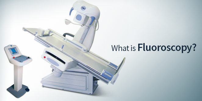
Fluoroscopy is a form of medical imaging that uses a series of X-rays to show the inside of your body in real time, like a video. Healthcare providers use it to diagnose conditions and to help guide medical procedures. Common examples of fluoroscopy include angiography, barium swallow, cardiac catheterization and stent or catheter placement.
What is fluoroscopy?
Fluoroscopy is a medical imaging procedure that uses X-rays to show internal organs and tissues working in real time. Providers use fluoroscopy to diagnose issues with your organs or help guide them while performing medical procedures.
Compared to plain X-rays, which get a snapshot of a part of your body, fluoroscopy gets images of what your body’s doing as it happens. Think of it like the difference between a still photograph of a moment in time versus recording an event so you can see how it unfolds.
Types of Fluoroscopy
Musculoskeletal Fluoroscopy
What the procedure does: Allows for exact injections that treat chronic or acute pain
Procedure length: 5-10 minutes
Barium Swallow
What the scan evaluates: Your esophagus; a "modified" swallow looks at your swallowing function
The scan involves: Drinking x-ray dye while x-ray images are being taken of your throat and chest. You will be asked to move into different positions to take these x-ray images. You may be asked to swallow different forms of x-ray dye.
Prep: Do not eat, drink, chew or smoke anything after midnight the night before
Procedure length: 30 minutes
Fluoroscopic Enteroclysis
What the scan does: Evaluates your small intestine
The scan involves: Having a small tube placed into your nose and through your esophagus and stomach into your small intestine
Procedure length: 2-4 hours
Afterwards: You feel full or bloated and cramping may occur
Fluoroscopic Defecography
What the scan evaluates: Your rectum
The scan involves: A small tube inserted 1-2 inches into your rectum
Prep: Bowel cleansing
Procedure length: 30-60 minutes
Fluoroscopic Small Bowel Follow Through
The scan evaluates: Your small intestine
The scan involves: X-ray images will be taken of your abdomen until the x-ray dye travels all the way through your small intestine
Prep: Do not eat, drink, chew or smoke anything after midnight the night before your scheduled procedure
Procedure length: 2-4 hours
Afterward: You may feel full or bloated and cramping may occur
Fluoroscopic IVP (Intravenous Pyelogram)
The scan evaluates: Your urinary tract
The scan involves: X-ray dye injected into a vein in your arm or hand; images taken of your kidneys, ureters and bladder as they fill with the x-ray dye
Procedure length: 60 minutes
Prep: Bowel cleansing preparation starting no later than 12 noon on the day before
A Fluoroscopic VCUG (voiding cystourethrogram)
The scan evaluates: Your bladder and lower urinary tract
The scan involves: A small tube inserted into your bladder
Procedure length: 30-60 minutes
Fluoroscopic HSG (hysterosalpingogram)
The scan evaluates: Your uterus and fallopian tubes
The scan involves: A speculum placed into your vagina so that a small tube can be placed into your uterus. Once the tube is in place, the speculum will be removed. X-ray dye will be injected through that tube and fill your uterus and fallopian tubes. This may cause some cramping.
Procedure length: 30 minutes
What is fluoroscopy mainly used for?
Healthcare providers use fluoroscopy to help diagnose issues with specific body parts. They can also use it to help them guide procedures (like placing medical devices) inside your body or during injection or aspiration (known as interventional guidance).
Diagnostic fluoroscopy
Healthcare providers can use fluoroscopy to diagnose conditions in many parts of your body. Some examples include:
- Angiography. Providers use an angiogram to identify and diagnose narrowing or blockages in your arteries.
- Barium swallow (esophagogram). This test checks for problems in your upper gastrointestinal (GI) tract.
- Barium enema. This test checks for problems in your colon and rectum (parts of your large intestine).
- Cystography. Providers use cystography to help diagnose problems in your bladder. Voiding cystourethrogram (VCUG) is one type of cystography that checks how well your bladder drains.
- Hysterosalpingogram. In this procedure, a provider gets images of your uterus and fallopian tubes.
- Myelography. Myelography gets images of your spinal cord, nerve roots and spinal lining (meninges).
- Sniff test. This test checks how well your diaphragm is working.
Fluoroscopy for procedure guidance
Providers can use fluoroscopy during procedures to help them guide surgical instruments or medical devices. Examples include:
- Intravascular catheterization. In this procedure, fluoroscopy helps your provider see blood flowing through your arteries.
- Catheter insertion or adjustment. Fluoroscopy can help providers properly place catheters — thin tubes that help get fluids into your body or drain fluids from your body. Providers can place catheters through your urethra, blood vessels and bile ducts. For instance, fluoroscopy is often used in angioplasty procedures.
- Placement of stents. Fluoroscopy can help providers place stents, which are devices that help open narrow or blocked blood vessels.
- Orthopedic surgery. Your surgeon may use fluoroscopy to help guide orthopedic procedures, like joint replacement and fracture (broken bone) repair.
How does fluoroscopy work?
Fluoroscopy works by using a special camera that uses pulses (brief bursts) of X-ray beams to take pictures of your insides. This can be while your organs perform their normal tasks or while your provider performs a procedure.
Some procedures use a contrast agent to help the provider see your organs and structures better. You might hear it called a dye, but it isn’t the type of dye that can stain your clothes. Your provider might:
- Inject the dye into your vein
- Have you drink a liquid with the dye in it
- Apply the dye inside your rectum with an enema
How do I prepare for fluoroscopy?
Your preparation will depend on the type of fluoroscopy procedure and why you’re getting it. Some procedures don’t require any special preparations. For others, your provider may have you avoid certain medications and/or fast (not eat or drink anything) for several hours before the procedure. Ask your provider if you have questions about what you need to do before the procedure.
- Precautions: If you are pregnant or think you may be pregnant, please check with your doctor before scheduling the exam. Other options will be discussed with you and your doctor.
- Clothing: You may be asked to change into a patient gown. A gown will be provided for you. Lockers are provided to secure your personal belongings. Please remove all piercings and leave all jewelry and valuables at home.
- Eat/Drink: Specific instructions will be provided based on the examination you are scheduled for.
- Allergies: Notify the radiologist or technologist if you are allergic or sensitive to medications, contrast dyes or iodine.
What happens during a fluoroscopy test?
Depending on the type of procedure, you may have your fluoroscopy at an outpatient center or as part of your stay in a hospital. Right before the procedure, you may need to remove jewelry or change into a gown.
Your fluoroscopy may include the following steps:
- You’ll lay on a table or sit in a chair.
- An anesthesiologist will give you general anesthesia through a vein in your arm (if you’ll be asleep for the procedure).
- You’ll swallow contrast dye or a provider will give it to you with an injection or enema (if it’s part of the procedure).
- Depending on the type of procedure, your provider may ask you to move your body into different positions. They may also ask you to hold your breath for a short time.
- If your procedure involves getting a catheter, your provider will insert a needle in the appropriate body part. This may be your groin, elbow or another area.
- Your provider will use an X-ray scanner to take fluoroscopic images, which they’ll view on a computer screen.
Are you awake during this test?
Depending on the test, you might be awake or sedated (under anesthesia) for a fluoroscopic procedure. For instance, you’re more likely to be sedated if your provider is using it as imaging guidance during surgery or stent placement.
Other fluoroscopy tests are mostly painless and might require you to be awake during the procedure so that you can follow your provider’s instructions. Your provider will let you know if you’ll have anesthesia for your procedure or not.
What are the risks of fluoroscopy?
The main risk of fluoroscopy is radiation exposure. Fluoroscopy for diagnostic purposes uses very low levels of radiation. When providers use fluoroscopy during a surgery, you’re exposed to radiation for a longer period of time, which could:
- Damage your skin and underlying tissues (“burns”)
- Increase your risk of cancer later in life
- Cause damage to the fetus if you’re pregnant
The likelihood of experiencing these side effects is very small. If the procedure is medically necessary, the benefit outweighs the possible radiation risks.
If contrast dye is part of your fluoroscopy procedure, there’s also a small risk of an allergic reaction. Be sure to tell your provider if you have any allergies or if you’ve ever had a reaction to contrast material.
X-Ray Dyes
Fluoroscopy procedures require various types of X-ray dyes, the choice of which depends upon the reason for the procedure. These dyes cause a selected part of the body to stand out from surrounding tissue in a scan. Some are thin like water, some thick like a milkshake, others carbonated like a soda, and still others are solid like a pill. Types of dyes used include:
- Barium sulfate, a white-chalky substance
- Water-soluble agents
- Omnipaque (iohexol)
- Hypaque (diatrizoic acid)
None of these dyes, or contrast agents, remain in your body on any kind of permanent basis.
What are the benefits of fluoroscopy?
The main benefit of fluoroscopy is that providers can see how your organs are working or where structures are while they perform a procedure. It’s unlike many other forms of medical imaging (like a CT scan) where you can only see one moment in time.
What is the difference between a CT scan and fluoroscopy?
Computed Tomography (CT) scans and fluoroscopy are two different imaging techniques used to diagnose medical conditions. A CT scan is a type of imaging that uses X-rays to create detailed 3D images of the body, while fluoroscopy is an imaging technique that uses X-rays to create real-time 2D images of the body. The main difference between CT scan and fluoroscopy is that CT is fully volumetric and better for low contrast imaging, such as imaging blood in the brain during stroke triage or changes in the gray and white matter which occur hours to days after a stroke. Modern CT scanners also do have real-time fluoroscopic imaging modes. However, traditional fluoroscopy has a higher frame rate and higher spatial resolution than CT and there are many clinical tasks where the higher frame-rate and spatial resolution of an x-ray fluoroscopy system outweigh the benefits of CT.
What type of results will I get after a fluoroscopy test?
The kind of results you get from a fluoroscopy test will depend on the kind of test you had. If the results show that a part of your body isn’t working as it should, you may need additional diagnostic tests or treatment. Ask your provider what you can expect.
When should I know the results?
Depending on the type of fluoroscopy test you’re having and whether or not it’s part of a surgical procedure, you might know the results of the test:
- While your provider is performing it or right after
- After you wake up from surgery
- After a radiologist or your specialist has reviewed the images, which takes anywhere from a day to about a week
Ask your provider when you can expect to know the results.
Is a fluoroscopy test painful?
Fluoroscopy imaging itself is painless and noninvasive. But if your healthcare provider is using fluoroscopy as imaging guidance during a procedure like surgery or intervention, you may experience pain due to the procedure, not the fluoroscopy. Your provider will let you know what kind of pain levels you can expect during and after your procedure.
One Final Note..
If you’ve ever taken your car to a mechanic, you know that they often can’t know what the issue is just by looking at a picture of your vehicle. They might have to watch it run and understand what’s happening as the car works. Fluoroscopy is a similar idea: Your provider needs to see your body working in order to know where the issue is and how to treat it. They can also use it to accurately place medical devices. Don’t hesitate to ask your provider about how the test works or what the results mean.
Find me on Social Media
 |
Don't forget to bookmark me to see updates.. Copyright © 2000 - 2025 - K. Kerr Most recent revision February 09, 2026 07:36:58 AM
|


