- Main Page
- A1C Test
- Advance Directives
- Anxiety
- Aortic Aneurysm
- Aphrodisiacs
- Apple Cider Vinegar
- Arrhythmia
- Atrial Fibrillation - AFib
- Back Pain
- Blood Cancer
- Blood Tests
- Blood Test Tubes
- Blood Types
- BMI Calculator
- Body Mass Index - BMI
- Bone Density Scan
- Bone Scan
- BPPV
- Bronchitis
- Cancer Terminology
- Carbohydrates
- Cardiac Catheterization
- Cardiovascular Disease
- Caregiver Glossary
- Caregiver Resources - LGBTQ+
- Caregiver Resources - MO
- Caregiver Resources - USA
- Continuous Glucose Monitors
- Cholesterol
- Citalopram
- COPD
- Coronary Artery Disease
- Cough
- CPAP
- CT scan
- Cyclobenzaprine
- Degenerative Disc Disease
- Depression
- Diabetes Information
- Diabetes - Type 1
- Diabetes - Type 2
- Diabetes - Type 3c
- Diabetes Facts
- Diabetes Care
- Diabetes Care Team
- Diabetes & Fruits
- Diabetes - Gestational
- Diabetes - Pre
- Diabetic Terms
- Diabetes & Vegetables
- Diet - Boiled Egg
- Diet - DASH
- Diet - Fat Burning
- Diet - Mediterranean
- Diet - Military
- Disability
- Disability Permits
- Do Not Resuscitate
- Dupixent®
- Echocardiogram
- E-Cigarettes
- Electrocardiogram
- Electromyography
- Emphysema
- Epidural - Lumbar
- Epidural - Transforaminal
- Epsom Salt
- Facet Arthropathy
- Farxiga®
- Flu - Influenza
- Fluoroscopy
- Gabapentin
- GERD
- Glycemic Index
- Gout
- Headaches
- Healing & Energy Work
- Health Facts
- Health Info. Lines
- Heart Attack
- Heart Disease - Other
- Heart Failure
- Heart Imaging Tests
- Herbal Terms
- Herbal Medicine
- Herb & Oils Uses
- Herniated disk
- HIPAA
- Home Remedies
- Humalog®
- Hydrogen Peroxide
- Hyperglycemia
- Hyperkalemia
- Hyperlipidemia
- Hypertension
- Hypoglycemia
- Hypokalemia
- Hypotension
- Important Numbers
- Indomethacin
- Informed Consent
- Inhalers
- Insomnia
- Insulin
- Juice Fasting
- Juice Recipes
- Kidney Cysts
- Kidney Disease
- Lantus®
- Lemon Benefits
- Lime Benefits
- Liver Disease
- Lumbar Retrolisthesis
- Lung Cancer
- Medicaid
- Medical Specialties
- Medicare
- Medicare - Your Rights
- Melatonin
- Men's Health
- Mental Health
- MO HealthNet
- Mounjaro®
- MRI Scan
- Multiple Myeloma
- Myelography
- Naproxen
- Nasal Polyps
- Nuclear Medicine
- Nutrition - Adults
- Nutrition - Adults, Older
- Nutrition - Kids
- Obesity
- Otolaryngologist
- Oxycodone-Acetaminophen
- Pain Management
- Peripheral Artery Disease
- Parking Spaces
- PET/CT Scan
- PET Scan
- Potassium
- Prednisone
- Prescription Drugs
- Prurigo Nodularis
- PVC's
- Quetiapine
- Quit Smoking
- Radiculopathy
- Red Yeast Rice
- Reiki
- Salt & Sodium
- Salt Water Flush
- Sciatica
- Service Animals
- Sleep Apnea
- Sleep Disorders
- Sleep Studies
- SPECT Scan
- Spinal Stenosis
- Statins
- Stents
- Stress Test - Exercise
- Stress Test - Nuclear
- Sugars - Sweeteners
- Support Groups
- Tardive Dyskinesia
- Testosterone
- Trazodone
- Ultrasound
- Vaccines 19 and up
- Vaccines by Age
- Vaccines 0-6 yrs
- Vaccines 7-18 yrs
- Ventricular Fibrillation
- Vertigo
- Vital Records
- Vital Signs
- Vitamin B12
- Vitamin C
- Vitamin D
- Vitamin E
- Vitamin F
- Vitamin K
- Vitamins and Minerals
- Vitamins Recommended
- Water Benefits
- X-Rays
Needed to read PDF's


Nuclear Cardiac Stress Test
Overview
The nuclear cardiac stress test is a key tool in heart health. It’s also known as myocardial perfusion imaging or radionuclide stress testing. This test checks blood flow to the heart muscle at rest and under stress.
This test helps doctors find heart areas that might not get enough blood. This is because of narrowed or blocked coronary arteries. It spots problems early, allowing for quick treatment and management of heart issues.
This guide will explain nuclear cardiac stress tests in detail. We’ll cover their purpose, how they’re done, what to expect, and what the results mean. If you’re thinking about this test or want to know more about it, keep reading. You’ll learn how it protects your heart health.
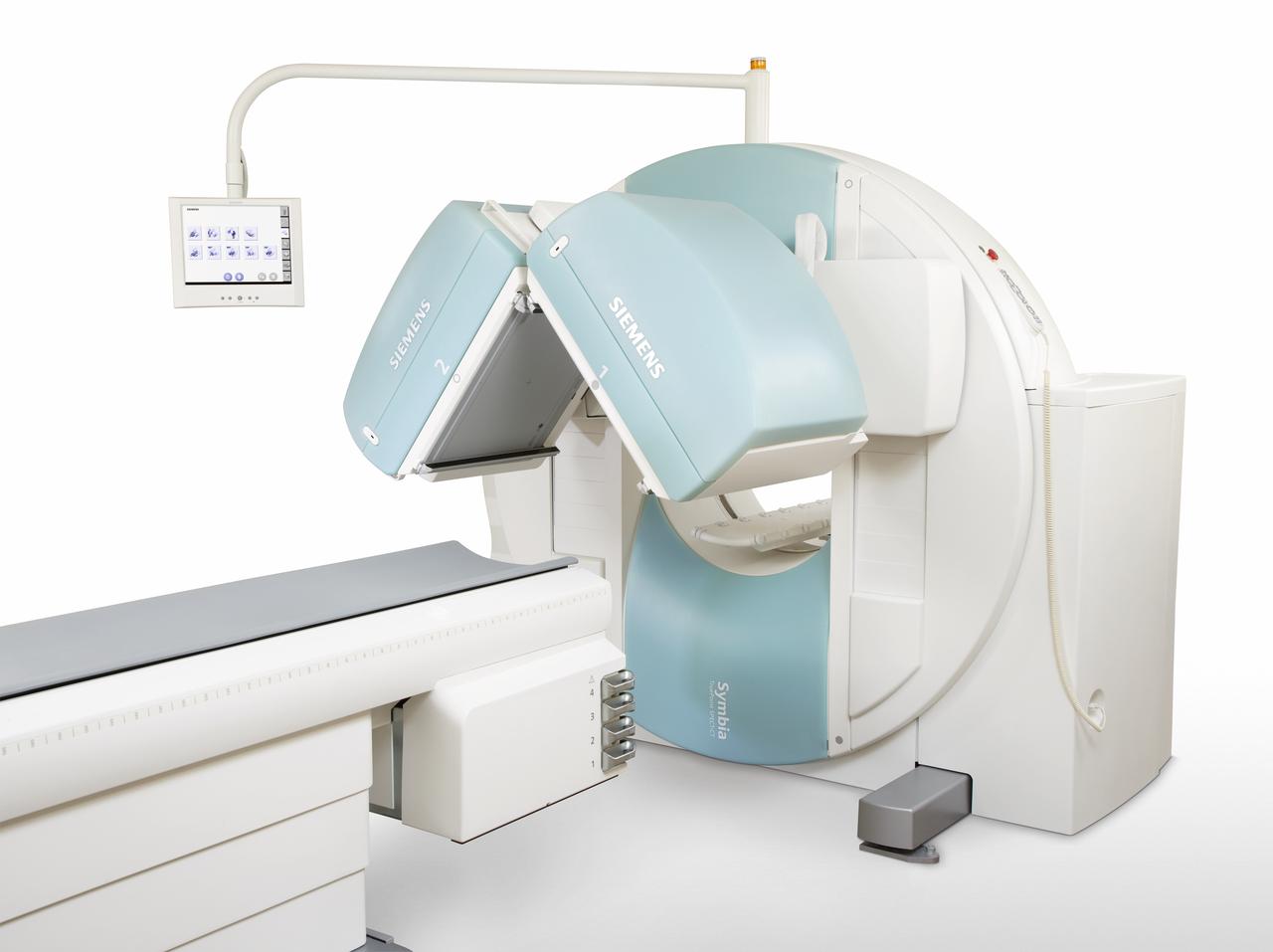
The test uses a positron emission technology (PET) scanner or single-photon emission computed tomography (SPECT) scanner.
Other names for a nuclear stress test are:
- Cardiac PET study (positron emission tomography)
- Cardiac SPECT study (single-photon emission computed tomography)
- Myocardial perfusion imaging (MPI) study.
What is a Nuclear Cardiac Stress Test?
A nuclear stress test, also known as a thallium stress test or cardiac nuclear imaging, checks how well the heart works. It looks at blood flow to the heart muscle at rest and when stressed. This test uses exercise or medicine to stress the heart and nuclear imaging to see the heart clearly.
This test mainly checks for coronary artery disease (CAD). CAD happens when heart arteries get narrow or blocked. It compares blood flow at rest and stress to find heart areas with less blood. This shows if there are blockages or damage.
How Nuclear Cardiac Stress Tests Differ from Other Cardiac Tests
Nuclear stress tests have big advantages over other heart tests like traditional stress tests or electrocardiograms (ECGs). The main differences are:
| Nuclear Stress Test | Traditional Stress Test |
| Provides images of blood flow to the heart | Measures electrical activity of the heart |
| Detects areas of reduced blood flow | Identifies abnormal heart rhythms |
| More sensitive in detecting CAD | May miss some cases of CAD |
Nuclear stress tests give detailed images of blood flow and heart function. This makes them better than other non-invasive tests for checking heart health. They are key in finding and managing coronary artery disease.
Indications for a Nuclear Cardiac Stress Test
Nuclear cardiac stress tests, also known as myocardial perfusion scintigraphy, are key in diagnosing and managing coronary artery disease. Doctors often suggest this test for several reasons:
Diagnosing Coronary Artery Disease
Patients with symptoms like chest pain, shortness of breath, or fatigue might need this test. It checks if the heart gets enough blood flow. By comparing heart images at rest and under stress, doctors spot areas with less blood flow. This shows possible blockages in the heart’s arteries.
Assessing the Severity of Known Coronary Artery Disease
For those with coronary artery disease, this test gives important details. It shows how much heart muscle is affected by poor blood flow. This helps doctors decide the best treatment.
Evaluating the Effectiveness of Treatment
This test also checks if treatments for coronary artery disease are working. By comparing images before and after treatment, doctors see if blood flow to the heart has improved. This helps them adjust the treatment plan if needed.
Whether or not to do a nuclear cardiac stress test depends on the patient’s situation. It looks at the heart’s function and coronary artery disease. This test is vital in guiding diagnosis and treatment plans.
Types of nuclear cardiac stress tests
Nuclear cardiac stress tests are key for finding and checking coronary artery disease. There are two main kinds: the exercise stress test and the pharmacological stress test. The right test depends on if the patient can exercise and their health.
Exercise Stress Test
An exercise stress test has the patient walk on a treadmill or bike while hooked up to an ECG. It watches how the heart acts when you’re active. This test is best for those who can exercise well and don’t have health issues.
This test shows how the heart works when you’re active. It looks at:
| Parameter | Normal Response | Abnormal Response |
| Heart Rate | Increases with exercise | Inadequate increase or excessive increase |
| Blood Pressure | Rises with exercise | Drops or fails to rise with exercise |
| ECG | Normal waveforms | ST-segment changes, arrhythmias |
| Symptoms | None | Chest pain, shortness of breath, dizziness |
Pharmacological Stress Test
A pharmacological stress test is for those who can’t exercise well. It uses a drug to make the heart work like it would during exercise. This is for people with health issues or on certain medicines.
Some drugs used are:
- Adenosine
- Dipyridamole
- Dobutamine
- Regadenoson
During the test, the heart’s rate, blood pressure, and ECG are watched closely. A special medicine is given to see how the heart works under stress.
What does a cardiac nuclear stress test show?
The nuclear stress test creates two images. The first shows how blood flows to your heart when you are resting. The second shows the blood flow during exercise. Cardiac stress testing is commonly used to diagnose and monitor coronary artery disease (CAD) by showing blood flow to your heart. CAD occurs when blood vessels are clogged or blocked.
The test can also:
- Determine whether your heart muscle is pumping well.
- Identify poor blood flow.
- The presence, location, and size of blockages that are serious enough to affect blood flow during exercise
- Show whether your heart has been damaged (for example, by a heart attack).
The images can tell your provider different things about your heart health:
- A normal test result will show blood being evenly distributed to the heart in both images.
- If both images show a fixed patch of poor blood flow, that means the person had a heart attack at some point.
- If an area of poor blood flow is seen on the exercise image, but not on the rest image, it means there is probably a blockage in one of the coronary arteries. The blockage is causing a temporary drop in blood flow.
It also may be used in people with:
- Acute coronary syndrome.
- Bundle branch block.
- Congestive heart failure.
- Other confirmed or suspected heart problems.
- Trouble breathing.
How Nuclear Cardiac Stress Tests Differ from Other Cardiac Tests
Nuclear stress tests have big advantages over other heart tests like traditional stress tests or electrocardiograms (ECGs). The main differences are:
| Nuclear Stress Test | Traditional Stress Test |
| Provides images of blood flow to the heart | Measures electrical activity of the heart |
| Detects areas of reduced blood flow | Identifies abnormal heart rhythms |
| More sensitive in detecting CAD | May miss some cases of CAD |
Nuclear stress tests give detailed images of blood flow and heart function. This makes them better than other non-invasive tests for checking heart health. They are key in finding and managing coronary artery disease.
Risks
Nuclear cardiac stress tests are usually safe but can have risks. These include radiation exposure, allergic reactions, and complications from exercise or medicine. Knowing these nuclear stress test risks helps patients make smart choices about their heart health.
Radiation Exposure
The main worry is radiation exposure. The test uses a radioactive tracer. The dose is low but not safe for pregnant women or those with certain health issues. Talk to your doctor about any radiation concerns.
Allergic Reactions to Radiopharmaceuticals
Some people might have allergic reactions to the test’s medicines. Symptoms include itching, hives, swelling, or trouble breathing. If you have allergies, tell your doctor before the test. Medical staff will watch for any allergic reactions.
Complications Related to Exercise or Pharmacological Stress
The test uses exercise or medicine to stress the heart. This can cause side effects like chest pain, shortness of breath, or dizziness. These effects are usually mild and go away quickly. But, serious problems like heart attack or stroke can rarely happen. If you have heart problems, talk to your doctor about the test’s risks and benefits.
Even with risks, nuclear cardiac stress tests are key for diagnosing heart disease. The benefits of early treatment often outweigh the risks. Work with your healthcare team to decide if the test is right for you, based on your health and risk factors.
How you prepare for a nuclear stress test?
Getting ready for a nuclear stress test is key to getting good results. By following the pre-test instructions and avoiding certain medicines, you help your healthcare team. They can then get the best info about your heart’s health.
Before your test, your doctor will give you specific instructions. These might include:
- Fasting for 4-6 hours before the test
- Avoiding caffeine for 24 hours before the procedure
- Wearing comfortable clothing and shoes suitable for exercise
- Bringing a list of your current medications
Medications to Avoid Before the Test
Some medicines can mess with the test’s accuracy. Your doctor might tell you to stop taking certain drugs before the test. Common ones to avoid include:
- Beta-blockers (e.g., metoprolol, atenolol)
- Calcium channel blockers (e.g., diltiazem, verapamil)
- Nitrates (e.g., nitroglycerin, isosorbide dinitrate)
- Dipyridamole (Persantine)
- Theophylline
Talking to your doctor about any medication changes is very important. They will give you advice based on your health and the medicines you take.
By following the right steps and listening to your doctor, you help make your test a success. This lets your healthcare team check your heart function well. They can then make a treatment plan that’s just right for you.
The Nuclear Cardiac Stress Test Procedure
A nuclear cardiac stress test, also known as myocardial perfusion imaging, is a non-invasive procedure. It checks blood flow to the heart muscle. The test has several steps.
First, a small amount of a radioactive tracer is injected into the bloodstream. This tracer builds up in the heart muscle based on blood flow. The patient then gets initial imaging while at rest, showing where the tracer is in the heart.
Next, the patient exercises, usually on a treadmill, to increase heart rate and stress the heart. If they can’t exercise, a pharmacological stress agent is given intravenously. At peak stress, a second tracer injection is given.
After the stress test, the patient gets a second imaging round. The images from rest and stress are compared. This helps find areas with less blood flow, which might show coronary artery blockages.
The whole nuclear cardiac stress test takes several hours, as shown in this timeline:
| Step | Duration |
| Initial injection and rest imaging | 30-60 minutes |
| Exercise or pharmacological stress | 10-20 minutes |
| Second injection and stress imaging | 15-30 minutes |
| Post-test monitoring | 30-60 minutes |
During the myocardial perfusion imaging, medical professionals closely watch the patient. While the test is generally safe, some patients may feel chest pain, shortness of breath, or dizziness during stress. These symptoms usually go away quickly after stopping the stress.
Interpreting Nuclear Cardiac Stress Test Results
Nuclear cardiac stress tests give important insights into your heart’s health. They compare images taken at rest and during stress. This helps doctors spot areas where blood flow is low, which might mean coronary artery disease.
It’s key for both patients and doctors to understand these results.
Normal vs. Abnormal Results
Normal tests show the radioactive tracer spreads evenly in the heart muscle at rest and stress. This means blood flow is good. But, abnormal results show spots where the tracer is low. This means some heart areas don’t get enough blood.
Abnormal tests can be broken down into a few types:
| Result | Interpretation |
| Fixed defect | Shows a spot with low tracer uptake at rest and stress. This might mean a heart attack or severe disease. |
| Reversible defect | Shows a spot with low uptake during stress but normal at rest. This suggests severe narrowing of a coronary artery. |
| Partial reversibility | Shows both fixed and reversible defects. This means a heart attack with more narrowing in arteries. |
Quantifying the Extent and Severity of Coronary Artery Disease
Nuclear stress tests can measure how bad coronary artery disease is. The size and where the defects are tell us about the arteries and blood flow. This helps decide the best treatment, like medicine or surgery.
These tests also calculate the left ventricular ejection fraction (LVEF). The LVEF is the heart’s pumping strength. A normal LVEF is 50% to 70%. A lower number might mean heart damage or weakness.
Benefits of Nuclear Cardiac Stress Testing
Nuclear cardiac stress testing is a powerful tool for diagnosing coronary artery disease. It shows detailed images of blood flow to the heart. This helps doctors see how well the heart is working and find any problems.
This test is great for catching coronary artery disease early. It lets doctors start treatment quickly, which can prevent bigger problems later. It also shows how bad the disease is, helping doctors choose the best treatment.
Here are some key benefits of nuclear cardiac stress tests:
| Advantage | Description |
| Accuracy | Provides detailed images of blood flow to the heart muscle |
| Early detection | Can identify coronary artery disease in its initial stages |
| Personalized treatment | Guides treatment decisions based on the severity of disease |
| Monitoring | Allows physicians to track the effectiveness of interventions over time |
Nuclear cardiac stress testing gives a full picture of heart health. It helps both patients and doctors make smart choices about treating coronary artery disease. The valuable insights from these tests can lead to quicker diagnosis, more focused treatment, and better health outcomes.
Alternatives to Nuclear Cardiac Stress Tests
Nuclear cardiac stress tests are useful, but there are other tests too. These tests can show how well the heart works and blood flows. They might be suggested based on a patient’s needs, medical history, and what they prefer.
Stress Echocardiography
Stress echocardiography uses ultrasound and stress, like exercise or medicine, to check the heart. It finds out if parts of the heart get enough blood when it needs it most. This test doesn’t use radiation, unlike some others.
Coronary Computed Tomography Angiography (CCTA)
Coronary computed tomography angiography (CCTA) makes detailed pictures of the heart and blood vessels with CT scans. It spots narrowed or blocked arteries without surgery. It’s good for people at low to moderate risk of heart disease.
Cardiac Magnetic Resonance Imaging (MRI)
Cardiac magnetic resonance imaging (MRI) uses magnets and radio waves to see the heart’s details. It shows the heart’s size, shape, and how it works. It also finds scar tissue or damage. MRI doesn’t use radiation and is a good choice for some patients.
The following table compares the advantages and disadvantages of these alternative tests:
| Advantages | Disadvantages |
| No radiation exposure, widely available, lower cost | Lower resolution images, operator-dependent |
| Non-invasive, high-resolution images, assesses coronary arteries | Radiation exposure, may require contrast dye |
| No radiation exposure, high-resolution images, assesses heart structure and function | Higher cost, longer scan times, not suitable for patients with certain implants |
Choosing between a nuclear cardiac stress test and other tests depends on many things. These include the patient’s condition, risk factors, and the doctor’s advice. Talking to a cardiologist can help pick the best test for each person.
Advancements in Nuclear Cardiac Stress Testing Technology
Nuclear cardiac stress tests have changed how doctors find and treat heart disease. The newest technology uses top-notch imaging and software. This gives doctors clear pictures of the heart.
These updates help spot blockages in coronary arteries better. They also check how well the heart works.
3D imaging is a big step forward. It shows the heart in more detail than 2D images. This helps doctors see problems more clearly.
New medicines are also being made. They help doctors see more without using too much radiation. This makes images clearer and safer for patients.
Technology has made these tests faster and easier for everyone. Patients now get through the test quicker and more comfortably. Doctors also have better tools to analyze the data.
As research keeps going, we can expect even better tests. These will help doctors diagnose and treat heart problems more accurately. This will be good for everyone’s heart health.
FAQ's
Here are answers to some common questions people ask about nuclear stress tests.
Q: Is a nuclear stress test painful?
A: A nuclear stress test is usually not painful. But some people may experience headaches, nausea, or dizziness after the test.
Q: How long are you radioactive after a nuclear stress test?
A: Depending on the type of radioactive tracer used, it may take a few hours to some days for it to leave your body through urine.
Q: Can you drive after nuclear stress test?
A: Some people may be able to drive after a nuclear stress test. But it may be safer to bring someone with you who can drive you back after the test.
Q: What should I avoid after a nuclear stress test?
A: Some people give off small amounts of radiation after a nuclear test. So, it will be best to avoid young children and babies after the test. Also, if you’re breastfeeding or chestfeeding, speak with your doctor about safety precautions.
Q: When will I get the results of a nuclear stress test?
A: It may take a few days for your healthcare team to process your test results, after which they will give you an appointment to discuss it with you.
Q: What is a nuclear cardiac stress test?
A: A nuclear cardiac stress test checks how well blood flows to the heart muscle. It uses a tiny bit of radioactive material and special cameras. This test is done at rest and during stress.
Q: Why would a doctor recommend a nuclear cardiac stress test?
A: A doctor might suggest this test to find coronary artery disease. It helps see how bad the disease is or if treatments are working.
Q: How should I prepare for a nuclear cardiac stress test?
A: To get ready, follow your doctor’s advice. This might mean fasting, avoiding caffeine and certain meds, and wearing comfy clothes and shoes for exercise.
Q: What happens during a nuclear cardiac stress test?
A: First, a tiny amount of radioactive material is injected into your blood. Then, you either walk on a treadmill or take a medication to simulate exercise. After that, your heart is scanned with special cameras.
Q: What are the different types of nuclear cardiac stress tests?
A: There are two main types. Exercise stress tests involve walking or biking. Pharmacological stress tests use medicine to mimic exercise.
Q: How are nuclear cardiac stress test results interpreted?
A: Results show how blood flows to the heart at rest and during stress. Normal results mean even blood flow. Abnormal results point to areas with less blood flow, showing the extent of heart disease.
Q: What are the risks and side effects of nuclear cardiac stress tests?
A: Risks include a small radiation dose and allergic reactions to the radioactive material. There might also be complications from exercise or the medication used.
Q: What are the benefits of nuclear cardiac stress testing?
A: This test accurately checks heart function and blood flow. It helps diagnose heart disease early. It also helps create personalized treatment plans.
Q: Are there any alternatives to nuclear cardiac stress tests?
A: Yes, there are alternatives. Stress echocardiography uses ultrasound. Coronary CT angiography uses CT scans. Cardiac MRI gives detailed heart images.
One Final Note..
A nuclear stress test is a noninvasive test used to show blood flow through the heart muscle during exercise and at rest. The test takes about 3 to 4 hours and usually doesn‘t cause serious complications. While this test is useful for diagnosing certain heart conditions, it may not be ideal for pregnant people.
A nuclear cardiac stress test helps diagnose and monitor heart problems such as coronary artery disease. A healthcare provider connects you to an EKG machine, injects a tracer into your bloodstream and takes images of blood flow to your heart before and after exercise. If you need a stress test, tell your doctor about any medications you take, and ask how you should prepare.
Find me on Social Media


 |
Don't forget to bookmark me to see updates.. Copyright © 2000 - 2025 - K. Kerr Most recent revision May 08, 2026 01:01:09 AM
|