- Main Page
- A1C Test
- Advance Directives
- Anxiety
- Aortic Aneurysm
- Aphrodisiacs
- Apple Cider Vinegar
- Arrhythmia
- Atrial Fibrillation - AFib
- Back Pain
- Blood Cancer
- Blood Tests
- Blood Test Tubes
- Blood Types
- BMI Calculator
- Body Mass Index - BMI
- Bone Density Scan
- Bone Scan
- BPPV
- Bronchitis
- Cancer Terminology
- Carbohydrates
- Cardiac Catheterization
- Cardiovascular Disease
- Caregiver Glossary
- Caregiver Resources - LGBTQ+
- Caregiver Resources - MO
- Caregiver Resources - USA
- Continuous Glucose Monitors
- Cholesterol
- Citalopram
- COPD
- Coronary Artery Disease
- Cough
- CPAP
- CT scan
- Cyclobenzaprine
- Degenerative Disc Disease
- Depression
- Diabetes Information
- Diabetes - Type 1
- Diabetes - Type 2
- Diabetes - Type 3c
- Diabetes Facts
- Diabetes Care
- Diabetes Care Team
- Diabetes & Fruits
- Diabetes - Gestational
- Diabetes - Pre
- Diabetic Terms
- Diabetes & Vegetables
- Diet - Boiled Egg
- Diet - DASH
- Diet - Fat Burning
- Diet - Mediterranean
- Diet - Military
- Disability
- Disability Permits
- Do Not Resuscitate
- Dupixent®
- Echocardiogram
- E-Cigarettes
- Electrocardiogram
- Electromyography
- Emphysema
- Epidural - Lumbar
- Epidural - Transforaminal
- Epsom Salt
- Facet Arthropathy
- Farxiga®
- Flu - Influenza
- Fluoroscopy
- Gabapentin
- GERD
- Glycemic Index
- Gout
- Headaches
- Healing & Energy Work
- Health Facts
- Health Info. Lines
- Heart Attack
- Heart Disease - Other
- Heart Failure
- Heart Imaging Tests
- Herbal Terms
- Herbal Medicine
- Herb & Oils Uses
- Herniated disk
- HIPAA
- Home Remedies
- Humalog®
- Hydrogen Peroxide
- Hyperglycemia
- Hyperkalemia
- Hyperlipidemia
- Hypertension
- Hypoglycemia
- Hypokalemia
- Hypotension
- Important Numbers
- Indomethacin
- Informed Consent
- Inhalers
- Insomnia
- Insulin
- Juice Fasting
- Juice Recipes
- Kidney Cysts
- Kidney Disease
- Lantus®
- Lemon Benefits
- Lime Benefits
- Liver Disease
- Lumbar Retrolisthesis
- Lung Cancer
- Medicaid
- Medical Specialties
- Medicare
- Medicare - Your Rights
- Melatonin
- Men's Health
- Mental Health
- MO HealthNet
- Mounjaro®
- MRI Scan
- Multiple Myeloma
- Myelography
- Naproxen
- Nasal Polyps
- Nuclear Medicine
- Nutrition - Adults
- Nutrition - Adults, Older
- Nutrition - Kids
- Obesity
- Otolaryngologist
- Oxycodone-Acetaminophen
- Pain Management
- Peripheral Artery Disease
- Parking Spaces
- PET/CT Scan
- PET Scan
- Potassium
- Prednisone
- Prescription Drugs
- Prurigo Nodularis
- PVC's
- Quetiapine
- Quit Smoking
- Radiculopathy
- Red Yeast Rice
- Reiki
- Salt & Sodium
- Salt Water Flush
- Sciatica
- Service Animals
- Sleep Apnea
- Sleep Disorders
- Sleep Studies
- SPECT Scan
- Spinal Stenosis
- Statins
- Stents
- Stress Test - Exercise
- Stress Test - Nuclear
- Sugars - Sweeteners
- Support Groups
- Tardive Dyskinesia
- Testosterone
- Trazodone
- Ultrasound
- Vaccines 19 and up
- Vaccines by Age
- Vaccines 0-6 yrs
- Vaccines 7-18 yrs
- Ventricular Fibrillation
- Vertigo
- Vital Records
- Vital Signs
- Vitamin B12
- Vitamin C
- Vitamin D
- Vitamin E
- Vitamin F
- Vitamin K
- Vitamins and Minerals
- Vitamins Recommended
- Water Benefits
- X-Rays
Needed to read PDF's


PET/CT
Overview
Positron emission tomography (PET) uses small amounts of radioactive materials called radiotracers or radiopharmaceuticals, a special camera and a computer to evaluate organ and tissue functions. By identifying changes at the cellular level, PET may detect the early onset of disease before other imaging tests can.
Tell your doctor if there is any possibility you are pregnant or you are breastfeeding. Your doctor will tell you how to prepare based on the type of your exam. Discuss any recent illnesses, medical conditions, medications you are taking and allergies – especially to contrast material. Your doctor will likely tell you not to eat anything and to drink only water for several hours before your scan. Leave jewelry at home and wear loose, comfortable clothing. You may wear a gown during the exam.
What is PET/CT scanning?
Positron emission tomography, also called PET imaging or a PET scan, is a type of nuclear medicine imaging.
Nuclear medicine uses small amounts of radioactive material called radiotracers. Doctors use nuclear medicine to diagnose, evaluate, and treat various diseases. These include cancer, heart disease, gastrointestinal, endocrine, or neurological disorders, and other conditions. Nuclear medicine exams pinpoint molecular activity. This gives them the potential to find disease in its earliest stages. They can also show whether you are responding to treatment.
Nuclear medicine is noninvasive. Except for intravenous injections, it is usually painless. These tests use radioactive materials called radiopharmaceuticals or radiotracers to help diagnose and assess medical conditions.
Radiotracers are molecules linked to, or "labeled" with, a small amount of radioactive material. They accumulate in tumors or regions of inflammation. They can also bind to specific proteins in the body. The most common radiotracer is F-18 fluorodeoxyglucose (FDG), a molecule similar to glucose. Cancer cells are more metabolically active and may absorb glucose at a higher rate. This higher rate can be seen on PET scans. This allows your doctor to detect disease before it may be seen on other imaging tests. FDG is just one of many radiotracers in use or in development.
You will usually receive the radiotracer in an injection. Or you may swallow it or inhale it as a gas, depending on the exam. It accumulates in the area under examination. A special camera detects gamma ray emissions from the radiotracer. The camera and a computer produce pictures and supply molecular information.
Many imaging centers combine nuclear medicine images with computed tomography (CT) or magnetic resonance imaging (MRI) to produce special views. Doctors call this image fusion or co-registration. Image fusion allows the doctor to connect and interpret information from two different exams on one image. This leads to more precise information and a more exact diagnosis. Single photon emission CT/CT (SPECT/CT) and positron emission tomography/CT (PET/CT) units can perform both exams at the same time. PET/MRI is an emerging imaging technology. It is not currently available everywhere.
A PET scan measures important body functions, such as metabolism. It helps doctors evaluate how well organs and tissues are functioning.
CT imaging uses special x-ray equipment, and in some cases a contrast material, to produce multiple images of the inside of the body. A radiologist views and interprets these images on a computer monitor. CT imaging provides excellent anatomic information.
PET/CT combines 2 types of imaging tests:
- Positron emission tomography (PET)
- Computed tomography (CT)
PET scans use a small amount of radiation to see and measure activity inside your body. A CT scan uses multiple x-rays to put together a picture of the inside of your body.
A computer uses this information to create a 3-dimensional picture of the body part being scanned.
PET/CT scans are useful for the early diagnosis of cancer. We can see a tumor and how the cells that make up the tumor work together. This can help us to find out if it's in fact cancer. PET/CT scanning can spot the spread of cancer to other areas of your body.
PET/CT scans are also used to study disorders of the:
- Brain
- Endocrine system
- Heart
- Cancer
What are some common uses of the procedure?
Doctors perform PET and PET/CT scans to:
- detect cancer and/or make a diagnosis.
- determine whether a cancer has spread in the body.
- assess the effectiveness of treatment.
- determine if a cancer has returned after treatment.
- evaluate prognosis.
- assess tissue metabolism and viability.
- determine the effects of a heart attack myocardial infarction on areas of the heart.
- identify areas of the heart muscle that would benefit from angioplasty or coronary artery bypass surgery (in combination with a myocardial perfusion scan).
- evaluate brain abnormalities, such as tumors, memory disorders, seizures and other central nervous system disorders.
- map normal human brain and heart function.
How should I prepare for a PET and PET/CT scan?
You may wear a gown during the exam or be allowed to wear your own clothing.
Women should always tell their doctor and technologist if they are pregnant or breastfeeding.
Tell the doctor and your exam technologist about any medications you are taking, including Vitamins and herbal supplements. List any allergies, recent illnesses, and other medical conditions.
You will receive specific instructions based on the type of your PET scan. Diabetic patients will receive special instructions to prepare for this exam.
If you are breastfeeding at the time of the exam, ask your radiologist or doctor how to proceed. It may help to pump breast milk ahead of time and keep it on hand for use until the PET radiotracer and CT contrast material are no longer in your body.
Leave metal objects including jewelry, eyeglasses, dentures and hairpins at home as they may affect the CT images. You may need to remove hearing aids and removable dental work.
Generally, your doctor will tell you not to eat anything for several hours before a whole body PET/CT scan. Eating may alter the distribution of the PET tracer in your body and can lead to a suboptimal scan. This could require you to repeat the scan on another day, so following instructions regarding eating is very important. You should not drink any liquids containing sugars or calories for several hours before the scan. Instead, you are encouraged to drink water. If you are diabetic, your doctor may give you special instructions. Tell your doctor about all the medications you are taking. List any allergies, especially to contrast materials or iodine.
Your doctor will check for any conditions you may have that could increase the risk of receiving intravenous contrast material.
What Is a PET/CT Scan Like?
- An IV is placed in your arm.
- You'll be give a small amount of the radioactive material for the PET scan.
- Wait for about 60 minutes.
- You'll have to lie flat on your back on the machine's table.
- The table moves slowly through a large ring. You'll need to be still for about 15 minutes while the scan is completed.
The placement of the IV may give you some discomfort, but there should be no other pain involved.
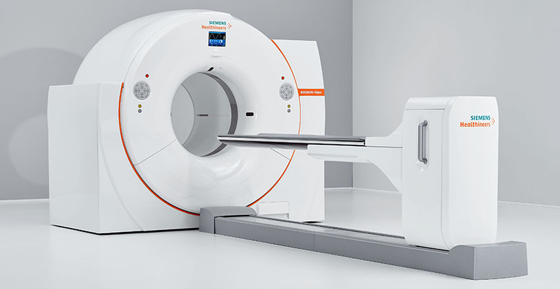
What does the equipment look like?
A PET scanner is a large machine with a round, donut-shaped hole in the middle. It looks like a CT or MRI unit. Multiple rings of detectors inside the machine record the energy emissions from the radiotracer in your body.
The CT scanner is typically a large, donut-shaped machine with a short tunnel in the center. You will lie on a narrow table that slides in and out of this short tunnel. Rotating around you, the x-ray tube and electronic x-ray detectors are located opposite each other in a ring, called a gantry. The computer workstation that processes the imaging information is in a separate control room. This is where the technologist operates the scanner and monitors your exam in direct visual contact. The technologist will be able to hear and talk to you using a speaker and microphone.
Combined PET/CT scanners look similar to both the PET and CT scanners.
A computer creates the images using the data from the gamma camera.
How does the procedure work?
Ordinary x-ray exams pass x-rays through the body to create an image. Nuclear medicine uses radioactive materials called radiopharmaceuticals or radiotracers. Your doctor typically injects this material into your bloodstream. Or you may swallow it or inhale it as a gas. The material accumulates in the area under examination, where it gives off gamma rays. Special cameras detect this energy and, with the help of a computer, create pictures that detail how your organs and tissues look and function.
PET scans only use radiotracer injections.
Unlike other imaging techniques, nuclear medicine focuses on processes within the body. These include rates of metabolism or levels of various other chemical activities. Areas of greater intensity are called “hot spots.” These may show large concentrations of the radiotracer and where there is a high level of chemical or metabolic activity. Less intense areas, or “cold spots,” indicate a smaller concentration of radiotracer and less activity.
How is the procedure performed?
Doctors perform nuclear medicine exams on outpatients and hospitalized patients.
You will lie on an exam table. If necessary, a nurse or technologist will insert an intravenous (IV) catheter into a vein in your hand or arm.
PET scans only use radiotracer injections.
The radiotracer typically takes about 30-60 minutes to travel through your body and be absorbed by the area under examination. You will be asked to rest quietly and to avoid movement and talking.
You may be asked to drink some contrast material that will localize in the intestines and help the radiologist interpreting the exam.
You will be moved into the PET/CT scanner to begin imaging. You will need to remain still during imaging. The CT exam takes place first, followed by the PET scan. On occasion, a second CT scan with intravenous contrast will follow the PET scan.
The CT scan takes less than two minutes. The PET scan takes 20-30 minutes.
Total scanning time is usually about 30 minutes.
Depending on which area is under examination, additional tests involving other tracers or drugs may be used. This could lengthen the procedure time to three hours. For example, if you are examined for heart disease, you may undergo a PET scan both before and after exercising or before and after receiving IV medication that increases blood flow to the heart.
After the exam, you may need to wait until the technologist determines if more images are needed. Sometimes, the technologist takes more images to clarify or better visualize certain areas or structures. The need for more images does not necessarily mean there was a problem with the exam or that something is abnormal. It should not cause you concern.
If you have an intravenous (IV) line for the procedure, your technologist will usually remove it. The technologist will leave it in place if you are to have another procedure that same day that requires an IV line.
What will I experience during and after the procedure?
During.
Except for intravenous injections, most nuclear medicine procedures are painless. Reports of significant discomfort or side effects are rare.
You will feel a slight pin prick when the technologist inserts the needle into your vein for the intravenous line. You may feel a cold sensation moving up your arm during the radiotracer injection. Generally, there are no other side effects.
PET scans only use radiotracer injections.
With some procedures, the technologist may place a catheter into your bladder. This may cause temporary discomfort.It is important to remain still during the exam. Nuclear imaging causes no pain. However, having to remain still or in one position for long periods may cause discomfort.
If you have a fear of closed spaces, you may feel anxious during the exam.
After
Unless otherwise directed, you may resume your normal routine and diet after the exam is completed. The radioactive tracer will remain in your body for a short time and will be excreted through the urine. You should drink plenty of fluids and empty your bladder frequently following your exam.
The small amount of radiotracer in your body will lose its radioactivity over time through the natural process of radioactive decay. It may also pass out of your body through your urine or stool during the first few hours or days after the test. Drink plenty of water to help flush the material out of your body.
Who interprets the results and how do I get them?
A radiologist or other doctor specially trained in nuclear medicine will interpret the images and send a report to your referring physician.
If your doctor has ordered a diagnostic CT, a radiologist with specialized training in interpreting CT exams will send a report to your referring physician.
What are the benefits vs. risks?
Benefits
- Nuclear medicine exams provide unique information that is often unattainable using other imaging procedures. This information may include details on the function and anatomy of body structures.
- Nuclear medicine supplies the most useful diagnostic or treatment information for many diseases.
- A nuclear medicine scan is less expensive and may yield more precise information than exploratory surgery.
- By identifying changes in the body at the cellular level, PET imaging may detect the early onset of disease before it is evident on other imaging tests such as CT or MRI.
The benefits of a combined PET/CT scan include:
- greater detail with a higher level of accuracy; because both scans are performed at the same time without the patient having to change positions, there is less room for error.
- greater convenience for the patient who undergoes CT and PET at one time rather than two different times.
Risks
- Because nuclear medicine exams use only a small dose of radiotracer, they have a relatively low radiation exposure. This is acceptable for diagnostic exams. Thus, the potential benefits of an exam outweigh the very low radiation risk.
- Doctors have been using nuclear medicine diagnostic procedures for more than six decades. There are no known long-term adverse effects from such low-dose exposure.
- Your doctor always weighs the benefits of nuclear medicine treatment against any risks. Your doctor will discuss the significant risks prior to treatment and give you an opportunity to ask questions.
- Allergic reactions to radiotracers are extremely rare and usually mild. Always tell the nuclear medicine personnel about any allergies you may have. Describe any problems you may have had during previous nuclear medicine exams.
- The radiotracer injection may cause slight pain and redness. This should rapidly resolve.
- Women should always tell their doctor and radiology technologist if there is any possibility that they are pregnant, or they are breastfeeding.
What are the limitations of PET/CT?
Nuclear medicine procedures can be time consuming. It can take several hours to days for the radiotracer to accumulate in the area of interest. Plus, imaging may take up to several hours to perform. In some cases, newer equipment can substantially shorten the procedure time.
The image resolution of nuclear medicine images may not be as high as that of CT or MRI. However, nuclear medicine scans are more sensitive for a variety of indications. The functional information they yield is often unobtainable using other imaging techniques.
Altered blood sugar or blood insulin levels may adversely affect the test results of diabetic patients or patients who have eaten a few hours prior to the exam.
The radiotracer decays quickly and is effective for only a short time. Therefore, it is important for you to be on time for your appointment and to receive the radioactive material at the scheduled time. Late arrival for an appointment may require you to reschedule the procedure.
A very obese person may not fit into the opening of a conventional PET/CT unit.
Find me on Social Media


 |
Don't forget to bookmark me to see updates.. Copyright © 2000 - 2025 - K. Kerr Most recent revision May 08, 2026 01:01:05 AM
|