- Main Page
- A1C Test
- Advance Directives
- Anxiety
- Aortic Aneurysm
- Aphrodisiacs
- Apple Cider Vinegar
- Arrhythmia
- Atrial Fibrillation - AFib
- Back Pain
- Blood Cancer
- Blood Tests
- Blood Test Tubes
- Blood Types
- BMI Calculator
- Body Mass Index - BMI
- Bone Density Scan
- Bone Scan
- BPPV
- Bronchitis
- Cancer Terminology
- Carbohydrates
- Cardiac Catheterization
- Cardiovascular Disease
- Caregiver Glossary
- Caregiver Resources - LGBTQ+
- Caregiver Resources - MO
- Caregiver Resources - USA
- Continuous Glucose Monitors
- Cholesterol
- Citalopram
- COPD
- Coronary Artery Disease
- Cough
- CPAP
- CT scan
- Cyclobenzaprine
- Degenerative Disc Disease
- Depression
- Diabetes Information
- Diabetes - Type 1
- Diabetes - Type 2
- Diabetes - Type 3c
- Diabetes Facts
- Diabetes Care
- Diabetes Care Team
- Diabetes & Fruits
- Diabetes - Gestational
- Diabetes - Pre
- Diabetic Terms
- Diabetes & Vegetables
- Diet - Boiled Egg
- Diet - DASH
- Diet - Fat Burning
- Diet - Mediterranean
- Diet - Military
- Disability
- Disability Permits
- Do Not Resuscitate
- Dupixent®
- Echocardiogram
- E-Cigarettes
- Electrocardiogram
- Electromyography
- Emphysema
- Epidural - Lumbar
- Epidural - Transforaminal
- Epsom Salt
- Facet Arthropathy
- Farxiga®
- Flu - Influenza
- Fluoroscopy
- Gabapentin
- GERD
- Glycemic Index
- Gout
- Headaches
- Healing & Energy Work
- Health Facts
- Health Info. Lines
- Heart Attack
- Heart Disease - Other
- Heart Failure
- Heart Imaging Tests
- Herbal Terms
- Herbal Medicine
- Herb & Oils Uses
- Herniated disk
- HIPAA
- Home Remedies
- Humalog®
- Hydrogen Peroxide
- Hyperglycemia
- Hyperkalemia
- Hyperlipidemia
- Hypertension
- Hypoglycemia
- Hypokalemia
- Hypotension
- Important Numbers
- Indomethacin
- Informed Consent
- Inhalers
- Insomnia
- Insulin
- Juice Fasting
- Juice Recipes
- Kidney Cysts
- Kidney Disease
- Lantus®
- Lemon Benefits
- Lime Benefits
- Liver Disease
- Lumbar Retrolisthesis
- Lung Cancer
- Medicaid
- Medical Specialties
- Medicare
- Medicare - Your Rights
- Melatonin
- Men's Health
- Mental Health
- MO HealthNet
- Mounjaro®
- MRI Scan
- Multiple Myeloma
- Myelography
- Naproxen
- Nasal Polyps
- Nuclear Medicine
- Nutrition - Adults
- Nutrition - Adults, Older
- Nutrition - Kids
- Obesity
- Otolaryngologist
- Oxycodone-Acetaminophen
- Pain Management
- Peripheral Artery Disease
- Parking Spaces
- PET/CT Scan
- PET Scan
- Potassium
- Prednisone
- Prescription Drugs
- Prurigo Nodularis
- PVC's
- Quetiapine
- Quit Smoking
- Radiculopathy
- Red Yeast Rice
- Reiki
- Salt & Sodium
- Salt Water Flush
- Sciatica
- Service Animals
- Sleep Apnea
- Sleep Disorders
- Sleep Studies
- SPECT Scan
- Spinal Stenosis
- Statins
- Stents
- Stress Test - Exercise
- Stress Test - Nuclear
- Sugars - Sweeteners
- Support Groups
- Tardive Dyskinesia
- Testosterone
- Trazodone
- Ultrasound
- Vaccines 19 and up
- Vaccines by Age
- Vaccines 0-6 yrs
- Vaccines 7-18 yrs
- Ventricular Fibrillation
- Vertigo
- Vital Records
- Vital Signs
- Vitamin B12
- Vitamin C
- Vitamin D
- Vitamin E
- Vitamin F
- Vitamin K
- Vitamins and Minerals
- Vitamins Recommended
- Water Benefits
- X-Rays
Needed to read PDF's


Positron Emission Tomography
(PET)
Overview
A positron emission tomography (PET) scan is an imaging test that can help reveal the metabolic or biochemical function of your tissues and organs. The PET scan uses a radioactive drug called a tracer to show both typical and atypical metabolic activity. A PET scan can often detect the atypical metabolism of the tracer in diseases before the disease shows up on other imaging tests, such as computerized tomography (CT) and magnetic resonance imaging (MRI).
The tracer is most often injected into a vein within your hand or arm. The tracer will then collect into areas of your body that have higher levels of metabolic or biochemical activity. This often pinpoints the location of the disease. The PET images are typically combined with CT or MRI and are called PET-CT or PET-MRI scans.
What is a PET scan?
A positron emission tomography (PET) scan is an imaging test that produces images of your organs and tissues at work. The test uses a safe, injectable radioactive chemical called a radiotracer and a device called a PET scanner.
The scanner detects diseased cells that absorb large amounts of the radiotracer, which indicates a potential health problem.
Healthcare providers frequently use PET scans to help diagnose cancer and assess cancer treatment. They can also assess certain heart and brain issues with the scan.
What’s the difference between a PET scan, CT scan and MRI?
Definition and Basic Principles
PET scans use radioactive tracers to see how the body works. These tracers are injected into a vein. They collect in areas with high activity, creating 3D images of how well tissues and organs are working.
This makes PET scans great for finding and tracking diseases like cancer, heart disease, and brain disorders.
Differences Between PET and Other Imaging Techniques
CT and MRI scans show the body’s structure. But PET scans focus on how things are working at the cellular level. Here’s a quick comparison:
| Imaging Technique | Key Features | Best Used For |
| PET Scan | Uses radioactive tracers to show metabolic activity | Detecting cancer, heart problems, brain disorders |
| CT Scan | Uses X-rays to create detailed cross-sectional images | Diagnosing injuries, bone and joint problems, tumors |
| MRI Scan | Uses magnetic fields and radio waves to produce high-resolution images | Examining soft tissues, organs, and the central nervous system |
By using different imaging techniques together, doctors can understand a patient’s condition better. This leads to earlier diagnosis and more effective treatments.
Why it's done
A PET scan is an effective way to help discover a variety of conditions, including cancer, heart disease and brain disorders. Your health care provider can use this information to help diagnose, monitor or treat your condition.
Applications of PET Scans in Cancer Detection
PET scans have changed how we detect and manage cancer. They give us deep insights into how tumors work and grow. This helps doctors stage cancer accurately, track how treatments work, and spot cancer coming back. It’s all about making cancer care more personal and precise.
One big use of PET scans is in cancer staging. They show how far cancer has spread. This helps doctors choose the best treatment plan. Here’s a comparison of the TNM staging system and what PET scans can show:
| PET Scan Information | |
| T (Tumor) | Determines primary tumor size and extent of local invasion |
| N (Nodes) | Detects cancer spread to nearby lymph nodes |
| M (Metastasis) | Identifies distant metastases in organs and tissues |
PET scans are also key in checking how well treatments work. By comparing scans before and after treatment, doctors can see if it’s working. This helps them make changes to treatments to get better results and reduce side effects.
Also, PET scans help find cancer coming back early. They spot small, active cancer cells before symptoms show. This is very important for cancers like lung, breast, and colon cancer, which often come back.
PET scans are now a big part of precision medicine in cancer care. They work with genetic testing to make treatments fit each patient’s cancer. This makes treatments more effective and safer, leading to better health and life quality for patients.
PET Scan in Alzheimer’s and Neurological Disorders
PET scans are now a key tool in diagnosing and tracking Alzheimer’s disease and other neurodegenerative diseases. They help find amyloid plaques and tau tangles in the brain. This makes it easier to spot these brain disorders early and track how they change over time.
Early Diagnosis and Monitoring Disease Progression
PET scans are great for catching brain changes before symptoms show up. They spot amyloid plaques and tau tangles, signs of Alzheimer’s. This early catch is key for starting treatment right away.
They also help track how these diseases get worse. By comparing scans, doctors can see how fast the disease is growing. This helps them check if treatments are working and adjust plans as needed.
Heart disease
A positron emission tomography (PET) scan of the heart is an imaging test that uses specialized dye to allow your doctor to view problems with your heart.
The dye contains radioactive tracers, which concentrate on areas of the heart that may be injured or diseased. Using a PET scanner, your doctor can spot these areas of concern.
A heart PET scan is typically an outpatient procedure, meaning you will not have to stay at the hospital overnight. This is typically a same-day procedure.
Your doctor may order a heart PET scan if you’re experiencing symptoms of heart trouble.
Symptoms of heart trouble include:
- irregular heartbeat (arrhythmia)
- pain in your chest
- tightness in your chest
- trouble breathing
- weakness
- profuse sweating
Your doctor may also order a heart PET scan if other heart tests, such as an echocardiogram (ECG) or cardiac stress test, don’t provide your doctor with enough information. A heart PET scan can also be used to track the effectiveness of heart disease treatments.
Brain disorders
Glucose is the main fuel of the brain. During PET scans, tracers are “attached” to compounds such as glucose. By detecting radioactive glucose, the PET scan can show which areas of the brain are using glucose at the highest rates.
When a specialist interprets the scan, they can see how the brain is working and check for any irregularities.
PET scans are used to help diagnose and manage many CNS disorders, including:
- Alzheimer’s disease
- depression
- epilepsy
- head trauma
- Parkinson’s disease
Cardiovascular Evaluation with PET Scans
PET scans are a key tool for checking heart health and finding problems early. They show detailed images of blood flow and heart function. This helps doctors spot issues and plan the best treatments.
This non-invasive method is vital for spotting coronary artery disease. It’s a major cause of heart attacks and other heart problems.
Assessing Blood Flow and Heart Function
PET scans are great for checking how well blood flows to the heart. They use a special tracer to see where blood might not be reaching. This shows if there are blockages or narrow spots in the arteries.
They also look at how the heart uses energy. This is helpful for patients with heart failure or other heart issues. It helps doctors find parts of the heart that can get better with the right treatment.
Detecting Coronary Artery Disease
Coronary artery disease is a big risk to heart health. It happens when plaque builds up in heart arteries. PET scans are very good at finding this problem early.
They spot where blood flow or energy use is off. This helps doctors know where and how bad the problem is. Early detection is key to stopping the disease from getting worse.
With PET scans, doctors can create a treatment plan. This might include changing lifestyle habits, taking medicine, or surgery. Regular PET scans help see if treatments are working and make changes if needed.
When would I need a PET scan?
In general, a PET scan can measure vital functions, such as blood flow, oxygen use and blood sugar (glucose) metabolism. It can also identify organs and tissues that aren’t working as they should.
If your healthcare provider suspects you may have cancer, they’ll likely recommend a PET scan, which can detect cancer and/or make a diagnosis.
If you’ve already been diagnosed with cancer, your provider may recommend more than one PET scan throughout your treatment to:
- Determine whether the cancer has spread in your body (metastasized).
- Assess the effectiveness of treatment.
- Determine if the cancer has returned after treatment (recurred).
- Evaluate the prognosis (outlook) of the cancer.
If you’re having heart issues, your provider may recommend a PET scan to:
- Determine the effects of a heart attack on areas of your heart.
- Identify areas of the heart muscle that would benefit from angioplasty or coronary artery bypass surgery.
If you’re experiencing neurological symptoms, your provider may recommend a PET scan to evaluate possible brain abnormalities, such as tumors, seizures and other central nervous system conditions.
Test Details
How does a PET scan work?
A PET scan is a type of nuclear medicine imaging. Nuclear medicine uses small and safe amounts of radioactive material, called radiotracers, given through an IV.
Unlike other imaging techniques, PET scans focus on processes and molecular activity within your body. This gives them the potential to find disease in its earliest stages.
Diseased cells in your body absorb more of the radiotracer than healthy ones do. These are called “hot spots.” The PET scanner detects this radiation and produces images of the affected tissue. A PET/CT scan combines X-ray images from a CT scan with PET scan images.
Radioactive Tracers and Their Role
Before a PET scan, a small amount of radioactive tracer, like fluorodeoxyglucose (FDG), is injected into the patient. This tracer goes to cells that use a lot of glucose. Cancer cells, for instance, use more glucose than normal cells.
The Imaging Process
During the scan, the patient lies on a table that moves through a scanner. As the tracer decays, it releases positrons that hit nearby electrons, creating gamma rays. The PET scanner catches these rays and makes detailed 3D images of where the tracer is in the body.
Interpreting PET Scan Results
Radiologists look at PET scan images and calculate standardized uptake values (SUVs). SUVs show how much tracer a body part has taken in. This helps spot areas with unusual glucose use. Here’s a table showing SUV ranges for normal and cancerous tissues:
| Tissue Type | Typical SUV Range |
| Normal Tissue | 0.5 – 2.5 |
| Cancerous Tissue | 2.5 – 10.0+ |
By looking at PET images and SUVs, radiologists can find problems and give important health insights.
How do I prepare for a PET scan?
PET scans are an outpatient procedure, which means you go home the same day. Your healthcare provider will give you detailed instructions on how to prepare for the scan. In general, you should:
- Make sure your provider has a current list of all medications, Vitamins and supplements you take, as well as any allergies you have.
- Alert your provider if you think you could be pregnant or if you’re breastfeeding.
- Not eat anything for six hours before the test. Your healthcare provider may change this direction if you have diabetes.
- Drink only water.
- Avoid caffeine for 24 hours before the test if you’re being tested for a heart problem.
- Wear comfortable clothes and leave metal accessories, such as jewelry, eyeglasses, dentures and hairpins at home.
- Tell your healthcare provider if being in an enclosed space makes you anxious. You may be able to take a mild sedative to help you relax during the procedure.
What should I expect during a PET scan?
You can expect the following during a PET scan:
- You’ll receive an IV injection of a radiotracer that contains a safe amount of a radioactive drug. The most commonly used radiotracer is fluorodeoxyglucose (FDG).
- You’ll sit in a chair for about an hour while the radiotracer moves through your bloodstream and gets absorbed by your organs and tissues. Too much activity can send the radiotracer to areas of your body that your healthcare provider isn’t testing. You won’t be able to feel the radiotracer.
- If you’re getting a PET/CT scan, you may also get an IV injection of a contrast dye. This dye helps produce sharper CT images.
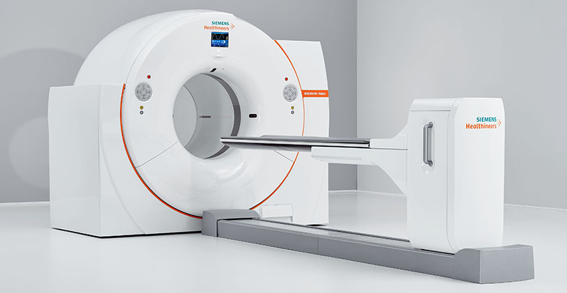
- You’ll lie on an exam table that slides in and out of the PET/CT scanner. This scanner is shaped like a doughnut. The doughnut or tunnel opening is about 30 inches in diameter.
- During the scan, which usually takes about 30 minutes, you must remain still. Movement can blur the images.
- You’ll hear buzzing and clicking sounds as the scanner takes images.
- A technologist will review the scans before you leave to ensure the images are in focus.
How long does a PET scan take?
The entire PET scan process takes about two hours.
It can take up to 60 minutes for your body to absorb the injected radiotracer. During this time, you’ll need to sit quietly and limit your movements. The actual PET scan takes about 30 minutes. After the test, you’ll need to wait while the technologist reviews the scans to ensure the images are clear.
What will I experience during and after the procedure?
Except for intravenous injections, most nuclear medicine procedures are painless. Reports of significant discomfort or side effects are rare.
You will feel a slight pin prick when the technologist inserts the needle into your vein for the intravenous line. You may feel a cold sensation moving up your arm during the radiotracer injection. Generally, there are no other side effects.
PET scans only use radiotracer injections.
With some procedures, the technologist may place a catheter into your bladder. This may cause temporary discomfort.
It is important to remain still during the exam. Nuclear imaging causes no pain. However, having to remain still or in one position for long periods may cause discomfort.
If you have a fear of closed spaces, you may feel anxious during the exam.
Unless your doctor tells you otherwise, you may resume your normal activities after your exam. A technologist, nurse, or doctor will provide you with any necessary special instructions before you leave.
The small amount of radiotracer in your body will lose its radioactivity over time through the natural process of radioactive decay. It may also pass out of your body through your urine or stool during the first few hours or days after the test. Drink plenty of water to help flush the material out of your body.
Results
A specialist trained to interpret scan images, called a radiologist, will report the findings to your provider. This process usually takes 24 hours.
The radiologist may compare your PET images with images from other tests you've undergone recently, such as an MRI or CT. Or the PET images may be combined to provide more detail about your condition.
Normal Results
A normal result means there were no problems seen in the size, shape, or position of an organ. There are no areas in which the tracer has abnormally collected.
What Abnormal Results Mean
Abnormal results depend on the part of the body being studied. Abnormal results may be due to:
- Cancer
- Infection
- Problem with organ function
Benefits vs. Risks
Benefits
- Nuclear medicine exams provide unique information that is often unattainable using other imaging procedures. This information may include details on the function and anatomy of body structures.
- Nuclear medicine supplies the most useful diagnostic or treatment information for many diseases.
- A nuclear medicine scan is less expensive and may yield more precise information than exploratory surgery.
- By identifying changes in the body at the cellular level, PET imaging may detect the early onset of disease before it is evident on other imaging tests such as CT or MRI.
The benefits of a combined PET/CT scan include:
- greater detail with a higher level of accuracy; because both scans are performed at the same time without the patient having to change positions, there is less room for error.
- greater convenience for the patient who undergoes CT and PET at one time rather than two different times.
Risks
- Because nuclear medicine exams use only a small dose of radiotracer, they have a relatively low radiation exposure. This is acceptable for diagnostic exams. Thus, the potential benefits of an exam outweigh the very low radiation risk.
- Doctors have been using nuclear medicine diagnostic procedures for more than six decades. There are no known long-term adverse effects from such low-dose exposure.
- Your doctor always weighs the benefits of nuclear medicine treatment against any risks. Your doctor will discuss the significant risks prior to treatment and give you an opportunity to ask questions.
- Allergic reactions to radiotracers are extremely rare and usually mild. Always tell the nuclear medicine personnel about any allergies you may have. Describe any problems you may have had during previous nuclear medicine exams.
- The radiotracer injection may cause slight pain and redness. This should rapidly resolve.
- Women should always tell their doctor and radiology technologist if there is any possibility that they are pregnant, or they are breastfeeding.
Considerations
Nuclear medicine procedures can be time consuming. It can take several hours to days for the radiotracer to accumulate in the area of interest. Plus, imaging may take up to several hours to perform. In some cases, newer equipment can substantially shorten the procedure time.
The image resolution of nuclear medicine images may not be as high as that of CT or MRI. However, nuclear medicine scans are more sensitive for a variety of indications. The functional information they yield is often unobtainable using other imaging techniques.
Altered blood sugar or blood insulin levels may adversely affect the test results of diabetic patients or patients who have eaten a few hours prior to the exam.
The radiotracer decays quickly and is effective for only a short time. Therefore, it is important for you to be on time for your appointment and to receive the radioactive material at the scheduled time. Late arrival for an appointment may require you to reschedule the procedure.
A very obese person may not fit into the opening of a conventional PET/CT unit.
Most PET scans are now performed along with a CT scan. This combination scan is called a PET/CT. This helps find the exact location of the tumor or other abnormality.
Research and Clinical Trials
PET scans are also big in research and trials for Alzheimer’s and other brain diseases. Scientists use them to study these diseases, find new treatments, and test new ideas.
They let researchers see and measure amyloid plaques and tau tangles in the brain. This helps them understand how these diseases work and find better treatments. Trials use PET scans to see if new treatments work and how patients react.
| Neurological Disorder | Role of PET Scan |
| Alzheimer’s Disease | Detects amyloid plaques and tau tangles for early diagnosis and monitoring progression |
| Parkinson’s Disease | Assesses dopamine function and monitors response to treatment |
| Huntington’s Disease | Evaluates brain metabolism and tracks disease progression |
PET Scan Safety and Radiation Exposure
PET scans are safe for most patients. The main worry is the radioactive tracers used. These tracers expose patients to low levels of radiation. But, the benefits of the scan usually outweigh the risks of low-dose exposure.
Doctors follow strict rules to keep radiation safety in check during PET scans. They control the radiation amount to keep it as low as needed. This ensures clear images without too much radiation.
Special care is needed for pregnant women and nursing mothers. Pregnant women should tell their doctor before a PET scan. The radiation could harm the fetus. Usually, safer imaging options like ultrasound or MRI are suggested instead.
Nursing mothers should talk to their healthcare provider too. They might be asked to pump and store breast milk before the scan. They should also avoid breastfeeding for a bit after to protect the baby from radiation.
Patients can feel safe knowing the medical team is trained in radiation safety. The equipment is kept in top shape for safe and accurate scans. This way, healthcare providers aim to get the most from PET scans while keeping risks low.
Advancements in PET Scan Technology
PET scanning has seen big changes in recent years. New technologies and the need for better diagnostic tools have driven these improvements. These changes have made PET scans more accurate and opened up new research areas.
Hybrid imaging systems, like PET/CT and PET/MRI scanners, are key breakthroughs. They combine PET’s metabolic info with CT or MRI’s detailed images. This gives doctors a full view of the body’s health, leading to better diagnoses and treatments.
Combined PET/CT and PET/MRI Scanners
PET/CT scanners are now central to molecular imaging. They mix PET and CT images perfectly. This helps find and understand diseases like cancer and heart issues more clearly.
| Advantage | PET/CT | Standalone PET or CT |
| Anatomical Localization | Precise fusion of functional and structural information | Limited anatomical context |
| Diagnostic Accuracy | Improved specificity and reduced false positives | Higher risk of misinterpretation |
| Treatment Planning | Enables targeted therapy and surgical guidance | Lacks detailed anatomical information for precise planning |
PET/MRI scanners also offer great benefits. They combine PET’s metabolic info with MRI’s detailed images. This is great for brain imaging and certain cancers, thanks to MRI’s soft tissue contrast.
Future Developments and Research
The future of PET scans looks bright. Researchers are working to make PET scans even better. They aim to improve how clear and accurate the images are. Artificial intelligence and machine learning will also change how we analyze PET images.
Personalized medicine is becoming more important, and PET scans will play a big role. They help tailor treatments to each patient’s needs. This could lead to better treatment results and fewer side effects.
As research advances, we can expect even more exciting changes in PET scan technology. New tracers and the integration of PET with other imaging methods will shape medical imaging. These developments will help diagnose and treat diseases better, benefiting patients everywhere.
Accessing PET Scan Services
To get a PET scan, you need a referral from your doctor. This involves checking your medical history and current health. This helps decide if a PET scan is right for you.
After getting a referral, your doctor will write a prescription. This prescription will say what kind of PET scan you need and any extra steps to follow.
Looking for a place to get your PET scan? Your doctor can recommend a good imaging center. You can also search online. Make sure the center is accredited by groups like the American College of Radiology. This means they follow strict quality and safety rules.
Insurance Coverage and Costs
How much insurance covers for PET scans depends on your plan and why you need it. Most plans, including Medicare and Medicaid, cover PET scans if they’re needed. But, you might have to pay a part of the cost, like a copay or deductible.
Check with your insurance to know what you’ll pay. If you don’t have insurance or it doesn’t cover much, some centers offer help. They might have lower prices or payment plans to make it easier to pay for the scan.
FAQ's
Q: What is a PET scan and how does it differ from other imaging techniques?
A: A PET scan uses radioactive tracers to see how the body works. It shows how cells use glucose, unlike CT or MRI scans. These scans mainly show the body’s structure, not how it functions.
Q: How do I prepare for a PET scan?
A: Before a PET scan, you might need to fast for hours and avoid exercise. Your doctor will tell you what to do, including any medicine changes. You’ll get a radioactive tracer through an IV and wait for about an hour before the scan starts.
Q: How is a PET scan used in cancer detection and management?
A: PET scans help doctors understand cancer by showing where it is and how it’s growing. They can spot cancer before it shows up on other scans. This helps doctors plan the best treatment for you.
Q: Can PET scans help diagnose Alzheimer’s and other neurological disorders?
A: Yes, PET scans are key in finding and tracking Alzheimer’s and other brain diseases. They can spot amyloid plaques and tau tangles in the brain. This helps doctors diagnose Alzheimer’s early and track how it progresses.
Q: Are PET scans safe, and how much radiation exposure is involved?
A: PET scans are mostly safe, with low radiation doses. The benefits of getting a clear diagnosis are worth the small risk. Pregnant and nursing women need extra care. Talk to your doctor about any concerns.
Q: How can I access PET scan services, and will my insurance cover the costs?
A: To get a PET scan, you need a doctor’s referral. They can find a good imaging center for you. Insurance for PET scans varies. Check with your provider to see what’s covered and what you might pay out of pocket.
One Final Note..
A positron emission tomography (PET) scan is a very useful and generally safe imaging test that healthcare providers use to assess cancer, heart issues and brain conditions. If you need a PET scan and are worried about the exam or have questions about it, don’t be afraid to ask your healthcare provider. They’re available to help and support you.
Find me on Social Media


 |
Don't forget to bookmark me to see updates.. Copyright © 2000 - 2025 - K. Kerr Most recent revision May 08, 2026 01:01:05 AM
|